
JOURNAL
Hydrodissection research for botulinum-toxin-related ptosis
Hydrodissection research for botulinum-toxin-related ptosis. Source observations, research context and limitations.
This article discusses the source author’s earlier literature, cases and experiences. Research populations and limitations matter; individual diagnosis and treatment require clinical assessment.
The source addresses persistent eyelid asymmetry reported four or five months after toxin treatment and the fear that it might be permanent.
It asks what has been reported for symptoms that persist rather than resolving as expected.
Most toxin-related ptosis improves as the effect wears off, but persistent symptoms require reassessment. The source introduces a 2026 technical protocol; a publication does not by itself establish efficacy.
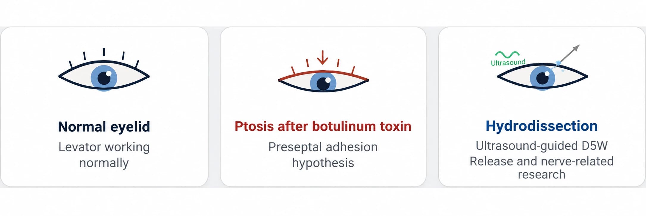
Natural recovery and persistent symptoms
Direct effects on the levator are one possible explanation. Persistent ptosis may have other causes, and it cannot be assigned to adhesions simply because several months have passed.
The hypothesis described by the paper
Lam KHS, Yoon Y, Hwang J, Yu H, et al. Ultrasound-guided preseptal hydrodissection with 5% dextrose in water for persistent botulinum toxin-associated ptosis: a technical protocol. Frontiers in Ophthalmology. 2026;6. https://doi.org/10.3389/fopht.2026.1792644
The source identifies Dr. Hyemi Yu and other collaborators and describes a clinical research process. Affiliations and roles are historical claims requiring verification. The paper proposes that the preseptal tissue environment may affect eyelid movement; this is a hypothesis, not a diagnosis for all persistent ptosis.
The proposed mechanism is that adhesions in the preseptal space could impede normal sliding. Persistent symptoms after expected toxin recovery do not independently confirm this mechanism.
A technical approach under investigation
The source introduces ultrasound-guided preseptal hydrodissection using 5% dextrose in water, or D5W. Evidence from peripheral nerve hydrodissection and a facial technical protocol must be distinguished. It is not presented as standard care or proof of comparative safety; technical instructions are not provided here.
The role of imaging
The eyelid contains closely related vessels, nerves and other structures. Imaging may support anatomical assessment but cannot eliminate procedural risks or establish superiority over another treatment.
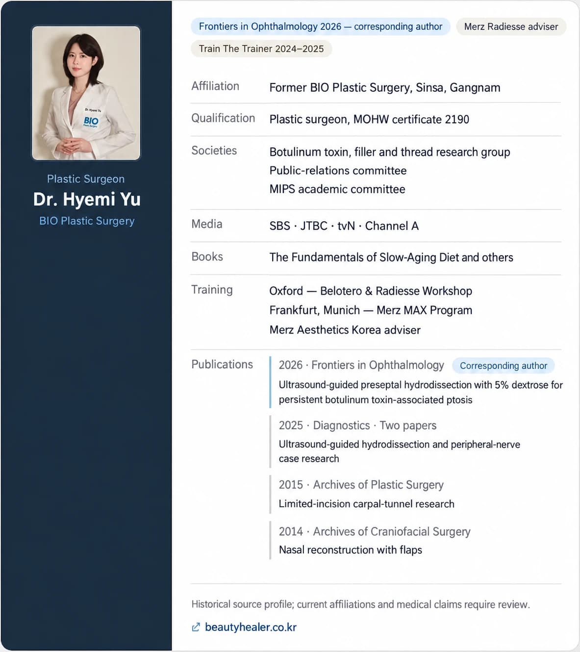
Persistent ptosis warrants a diagnosis before selecting treatment. Three months is a point discussed in the source, not a rule to wait before seeking evaluation. This literature discussion cannot determine an individual cause or appropriate procedure.
Discuss persistent or changing symptoms with an appropriately qualified clinician.
Source: Original post on Dr. Hyemi Yu’s Naver blog (Korean)
The original publication date and the website revision date are shown separately.