
JOURNAL
Fat calcification after facial surgery and fat grafting
Fat calcification after facial surgery and fat grafting. Source observations, research context and limitations.
This article discusses the source author’s earlier literature, cases and experiences. Research populations and limitations matter; individual diagnosis and treatment require clinical assessment.
The source describes people noticing a firm cheek lump or pulling during expression after a facelift combined with fat grafting. A surface irregularity may reflect several processes beneath the skin.
Source caption: an individual firm lesion after facelift and fat grafting.
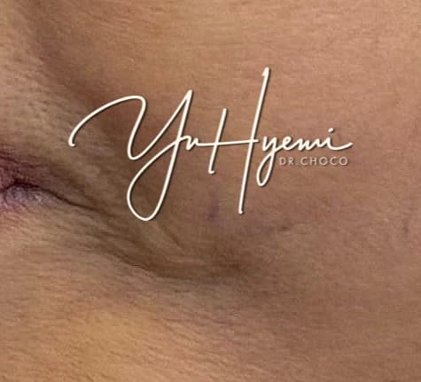
The patient in the source reported a palpable lump beside the cheek. Ultrasound was used to investigate its location and possible nature.
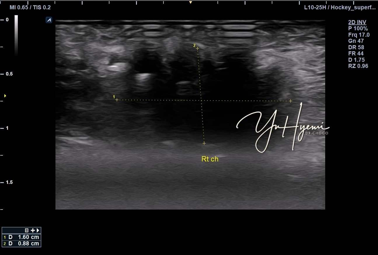
The author described a hypoechoic lesion in deeper subcutaneous fat, posterior acoustic shadowing and an irregular border without prominent acute inflammatory signs. Fat necrosis, fibrosis or partial calcification were considered; imaging impressions are not identical to a confirmed pathological diagnosis.
A facelift changes tissue planes and the graft recipient environment. Some grafted fat may not survive, and scar tissue or adhesions can affect movement. The source considered a conservative approach rather than automatically repeating surgery or fat grafting.
ESWT was selected in this individual case. The author discusses mechanical stimulation, remodeling and circulation using literature from other conditions. Such mechanisms do not establish a specific effect on every post-facial-grafting calcification. Ultrasound was repeated after three sessions.
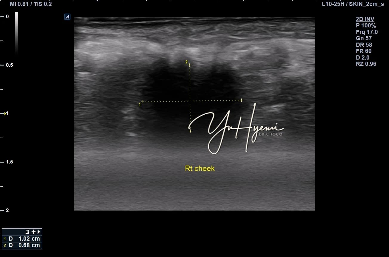
The maximum size remained around 1cm. The author felt the border and shadow appeared less pronounced and the tissue more homogeneous, while the patient reported less firmness and easier smiling. These subjective and imaging impressions were not a controlled outcome assessment; different imaging conditions can influence appearance.
The source discusses fibroblast activity, VEGF and microcirculation as possible mechanisms. It emphasizes that ESWT is not a method of instantly removing a calcified mass. Surgery and conservative management each have different indications and limits. The record illustrates one assessment and course, not a promised result or a reason to avoid surgery when it is needed.
Source: Original post on Dr. Hyemi Yu’s Naver blog (Korean)
The original publication date and the website revision date are shown separately.