
JOURNAL
Nodules and fat necrosis after HIFU
Nodules and fat necrosis after HIFU. Source observations, research context and limitations.
This article discusses the source author’s earlier literature, cases and experiences. Research populations and limitations matter; individual diagnosis and treatment require clinical assessment.
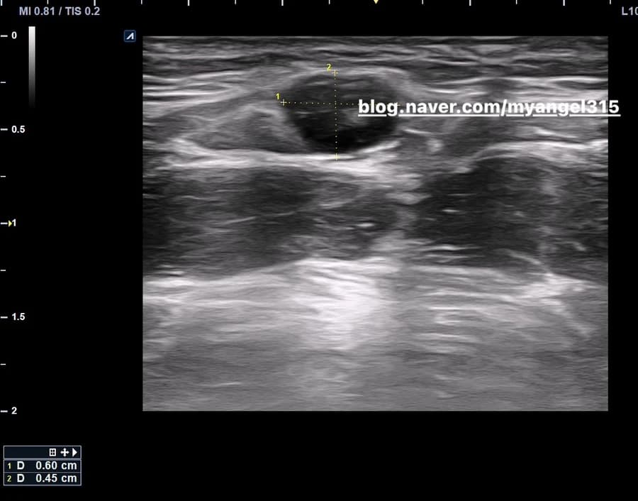
A clinical account of a nodule after HIFU
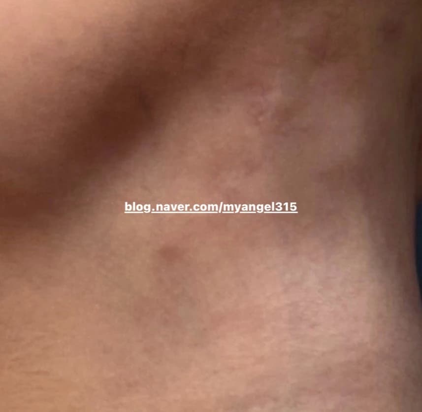
The source describes a palpable lesion of approximately 6mm after Shurink treatment, with a relatively defined hypoechoic area in superficial fat on ultrasound. The image is an observation, not a definitive diagnosis.
Heat, tissue and fluid
Shurink and Ultherapy are HIFU devices. The source discusses focal thermal coagulation at roughly 60–70°C and proposes that tissue thickness, fluid and local anesthetic infiltration can influence energy distribution. Its explanation that heat “stays in a space” is a simplified hypothesis requiring physical and clinical verification.
Fat injury can be followed by inflammation or fibrosis. A lesion’s cause and malignant potential cannot be established from this narrative alone. The source then discusses ESWT, mechanical tissue effects and circulation. These proposed mechanisms do not establish faster absorption in this patient.
Source caption: an immediate change in the lesion’s shape after ESWT.
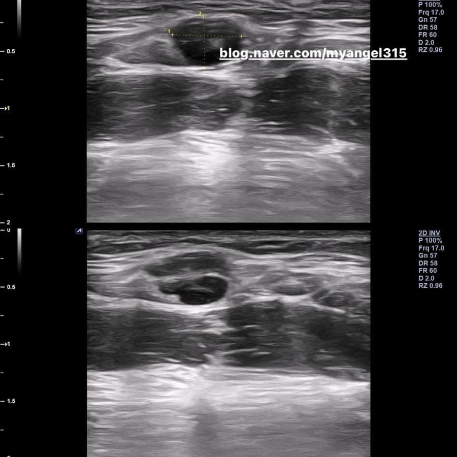
The source links musculoskeletal shock-wave research to aesthetic tissue remodeling. It observed a change in appearance, but several treatments were combined and the effect cannot be attributed to one intervention.
Combined approaches and their limits
The author discusses hydrodissection to separate tissue planes and a radiofrequency needle intended to produce controlled tissue effects. Peripheral nerve literature, facial nodules and individual combination outcomes are different questions. Neither improved circulation nor remodeling can be assumed solely from the proposed mechanism.
The source also recalls the author’s own nodule after Ultherapy and substantial anesthetic infiltration. That experience informed a preference for imaging and careful assessment rather than immediate excision; it does not establish a universal sequence or recovery rate.
The original reassurance that the lump was not cancer was removed: ultrasound impressions alone cannot exclude malignancy. Tissue depth and fluid conditions may affect treatment, but a 1–2mm analogy does not provide a general explanation or a safety guarantee.
Individual diagnoses, treatment choices and outcomes vary. Seek clinical assessment for a persistent or changing lesion.
Source: Original post on Dr. Hyemi Yu’s Naver blog (Korean)
The original publication date and the website revision date are shown separately.