
JOURNAL
Ultrasound and tissue assessment in nodule-treatment planning
Ultrasound and tissue assessment in nodule-treatment planning. An edited account of source experience, research context and limitations.
This edited account distinguishes personal experience, laboratory research and clinical studies. Individual suitability requires clinical assessment.
The source asks whether a palpable lump after filler or fat grafting should immediately be treated as something to remove, or first assessed to understand its nature. It discusses localized particles and redistribution, but nodules do not all have the same mechanism.
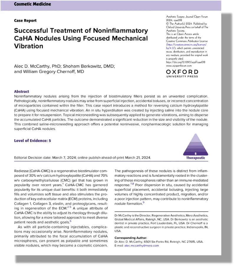
Pain, warmth, redness, timing, material and location matter. Absence of symptoms does not exclude inflammation or infection. CaHA, PDLLA and fat-grafting lumps can have different causes.
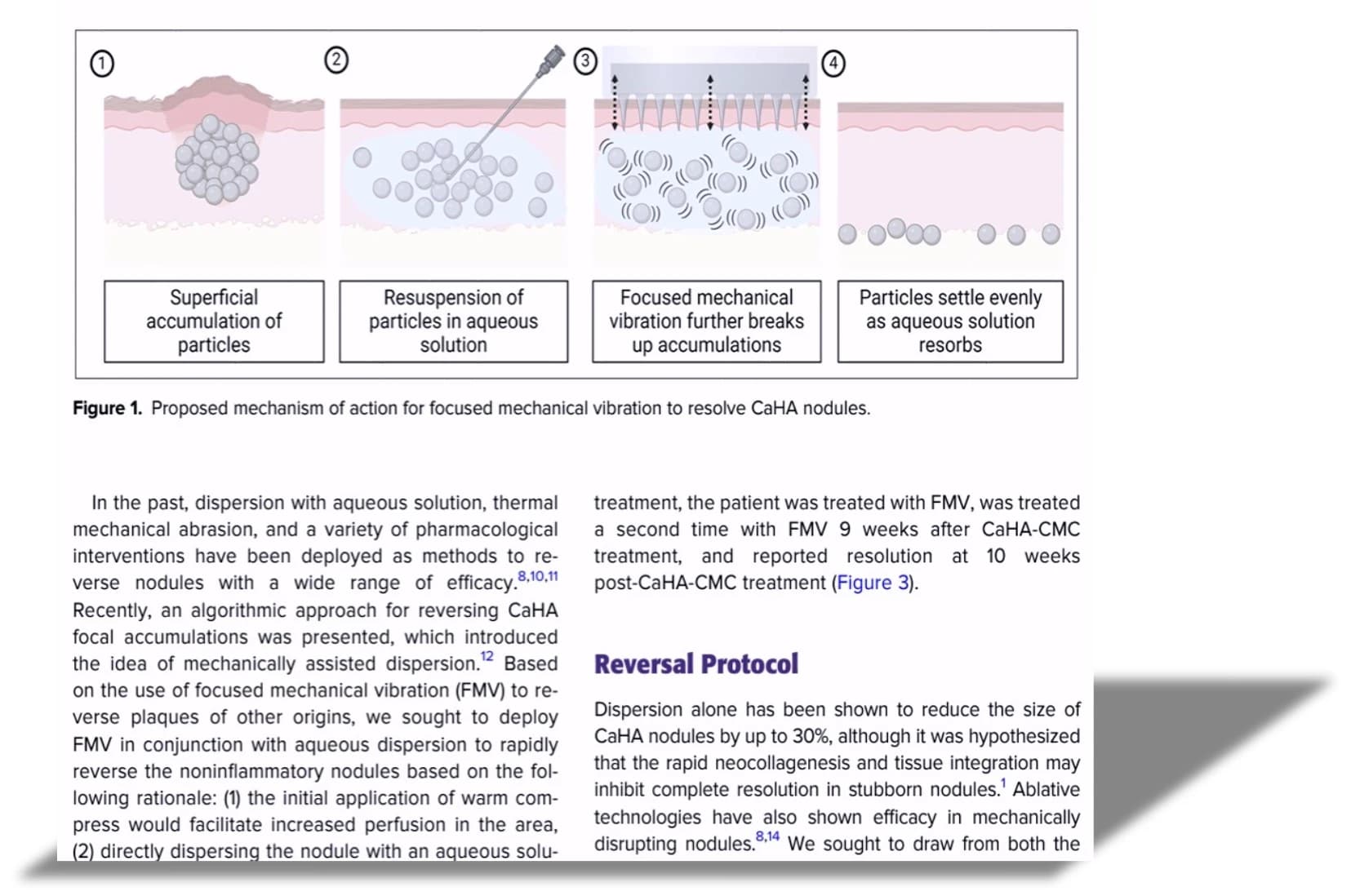
A source diagram illustrates a hypothesis about particles dispersing after fluid and mechanical stimulation. It is not proof that all tissue nodules behave that way. ESWT evidence also needs to be separated by material, diagnosis and study design.
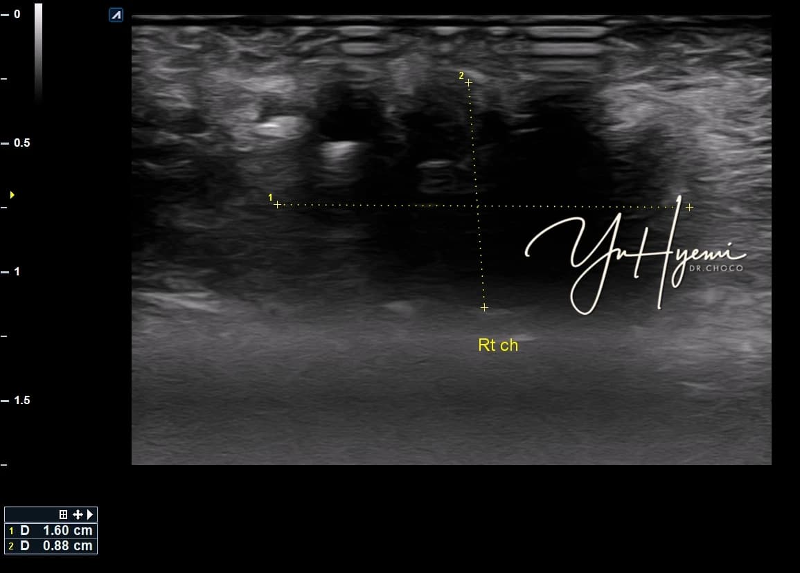
Changes in ultrasound size, borders or brightness can support follow-up. A hypoechoic area alone does not identify the material, and a smaller size does not establish cure.
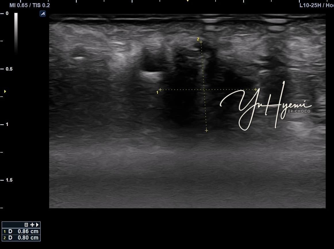
Strong pressure or self-massage is not presented as treatment. The article communicates a planning perspective combining imaging with symptoms and time. Unidentified papers and specific treatment evidence in the source still require confirmation.
Source: Original post on Dr. Hyemi Yu’s Naver blog (Korean)
The original publication date and the website revision date are shown separately.