
JOURNAL
Interpreting ultrasound follow-up of facial nodules
Interpreting ultrasound follow-up of facial nodules. An edited account of source experience, research context and limitations.
This edited account distinguishes personal experience, laboratory research and clinical studies. Individual suitability requires clinical assessment.
A facial nodule may be felt during touch or expression even when it is not easily visible. The source discusses ultrasound as a way to assess location, surrounding tissues and changes over time.
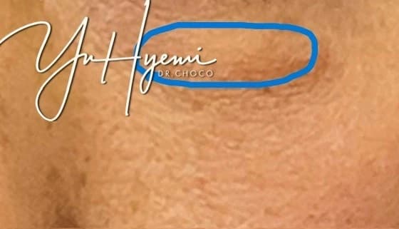
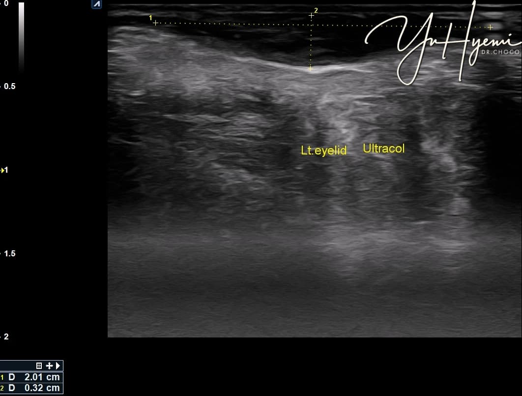
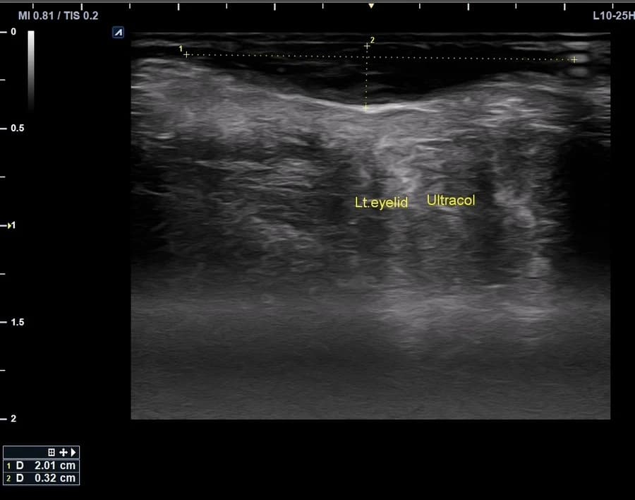
Brightness or posterior shadowing alone cannot establish the material, fibrosis or calcification. Findings vary with imaging settings, tissue structure and previous materials, so examination, symptoms and procedure records need to be considered together.
The author describes changing borders, distribution and firmness after ESWT, hydrodissection and other combined approaches. When several interventions overlap, their individual contribution is difficult to separate. The source’s combination is not presented as standard treatment for all nodules.
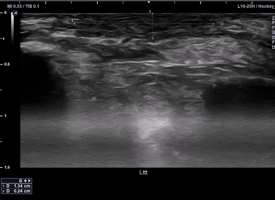
Symptoms may change without a major size difference, while a changed image does not mean treatment is complete. No fixed number or sequence is prescribed. This is an account of the limits of interpreting ultrasound follow-up.
Source: Original post on Dr. Hyemi Yu’s Naver blog (Korean)
The original publication date and the website revision date are shown separately.