
JOURNAL
Approaches to assessing under-eye fat nodules
Approaches to assessing under-eye fat nodules. Source observations, research context and limitations.
This article discusses the source author’s earlier literature, cases and experiences. Research populations and limitations matter; individual diagnosis and treatment require clinical assessment.
The under-eye region is thin and small changes may be noticeable. After fat grafting, some people report a protrusion or a firm area.
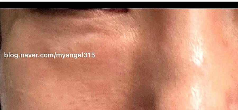
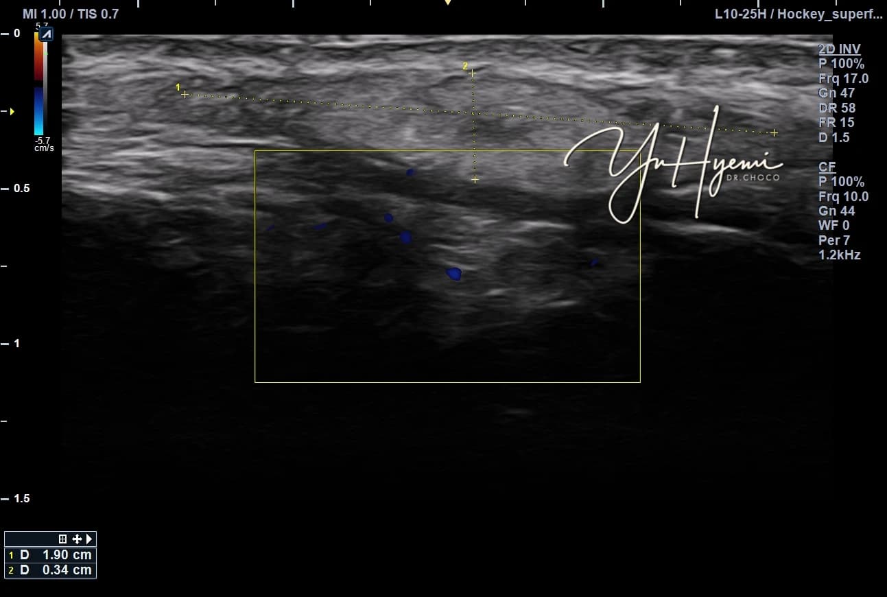
The source considers the graft recipient environment alongside volume. Tenderness may be limited even when a contour change is visible with makeup or smiling. The accompanying ultrasound relates to the individual photographed case.
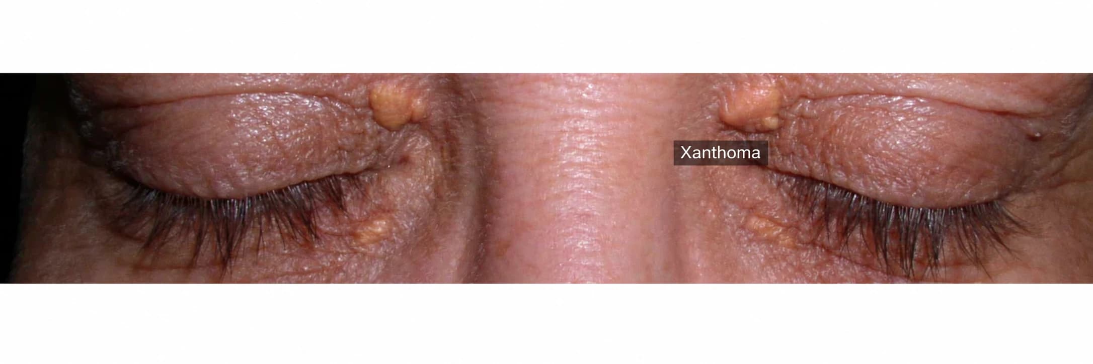
The source describes repeated lower-eyelid and xanthoma surgery before fat grafting, with thin tissue and adhesions. A later change was reported as less visible after treatment; this is not a general expected outcome.
Possible contributing factors
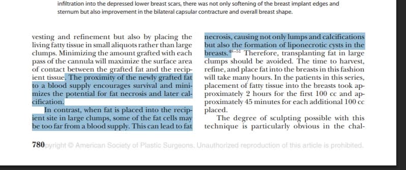
The source discusses vascular supply, scarring, pressure and fibrosis as influences on graft survival. Fat necrosis, fibrosis or calcification may follow in some cases. It cites Structural Fat Grafting: The Biological Basis, whose exact interpretation and applicability need comparison. Technique and recipient conditions should not be presented as mutually exclusive causes.
Is surgery always required?
The source cites Treatment Algorithm of Postsurgical Fat Necrosis in discussing conservative care when symptoms are limited and there is no rapid change or infection. The paper’s anatomical population and scope require checking; it is not a universal algorithm for every under-eye lesion. Surgery may be appropriate depending on the diagnosis and functional or cosmetic concern.
Goals of a conservative approach
The author discusses tissue stiffness, pressure and the surrounding environment, aiming to reduce symptoms rather than force complete removal. This perspective does not establish that an environmental treatment is superior to removal.
Ultrasound as part of assessment
Location, depth, relationship to fat and vessels and Doppler findings can contribute to an assessment. Ultrasound alone may not definitively distinguish necrosis, fibrosis or every cause of a lump.
The source emphasizes considering graft survival and surrounding tissue together. Responses can vary, and further treatment for a contour depression or thin skin needs its own assessment rather than a promise of recovery.
This is an individual clinical discussion, not guaranteed efficacy or an invitation to infer a diagnosis from photographs.
Source: Original post on Dr. Hyemi Yu’s Naver blog (Korean)
The original publication date and the website revision date are shown separately.