
JOURNAL
Ultrasound findings in collagen-booster nodules
Ultrasound findings in collagen-booster nodules. Source observations, research context and limitations.
This article discusses the source author’s earlier literature, cases and experiences. Research populations and limitations matter; individual diagnosis and treatment require clinical assessment.
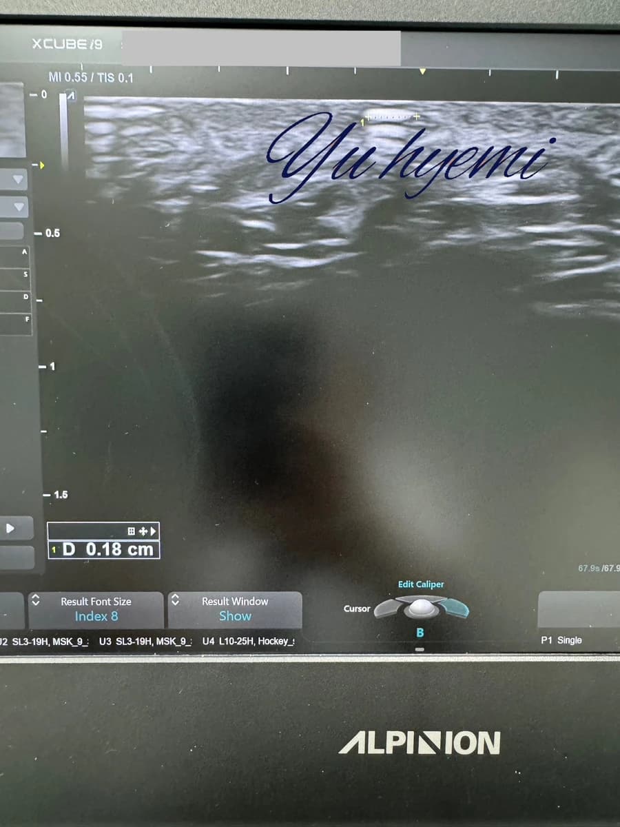
The source shows a bright area on ultrasound in an individual cheek CaHA case that was reportedly neither visible nor palpable at that point. It predicted disappearance within three to five months, but this is not a universal degradation or resolution timeline.
The original discusses CaHA particle sizes of 36–52μm, approximately 3μm surface features, changes described as around 40μm at one month and 20μm at seven months, and macrophage handling of smaller particles below 10μm. These numerical and histological claims require comparison with the underlying paper and cannot be inferred directly from this ultrasound.
The author considers superficial deposits and the original injection route as possible explanations. The source caption refers to a lesion noticed three weeks after treatment.
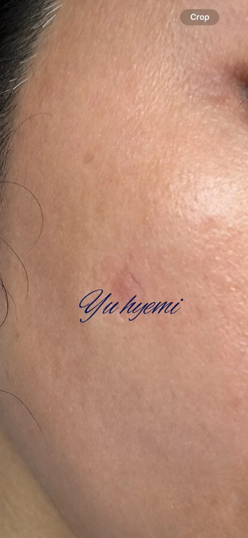
The author describes daily treatment over an early period and a lesion no longer visible or palpable after roughly two weeks, although it remained detectable on ultrasound. Pigmentation was discussed as a possible associated concern. The source’s two-week to three-month treatment range is a personal experience range, not a guarantee or general protocol.
Source: Original post on Dr. Hyemi Yu’s Naver blog (Korean)
The original publication date and the website revision date are shown separately.