
JOURNAL
Nodules after unknown injections: limits of ultrasound
Nodules after unknown injections: limits of ultrasound. Source observations, research context and limitations.
This article discusses the source author’s earlier literature, cases and experiences. Research populations and limitations matter; individual diagnosis and treatment require clinical assessment.
The source describes people with recurrent swelling, pulling or pain months or years after an injection whose contents they do not know. Symptoms and history matter more than a marketing name such as a “patented collagen injection.”
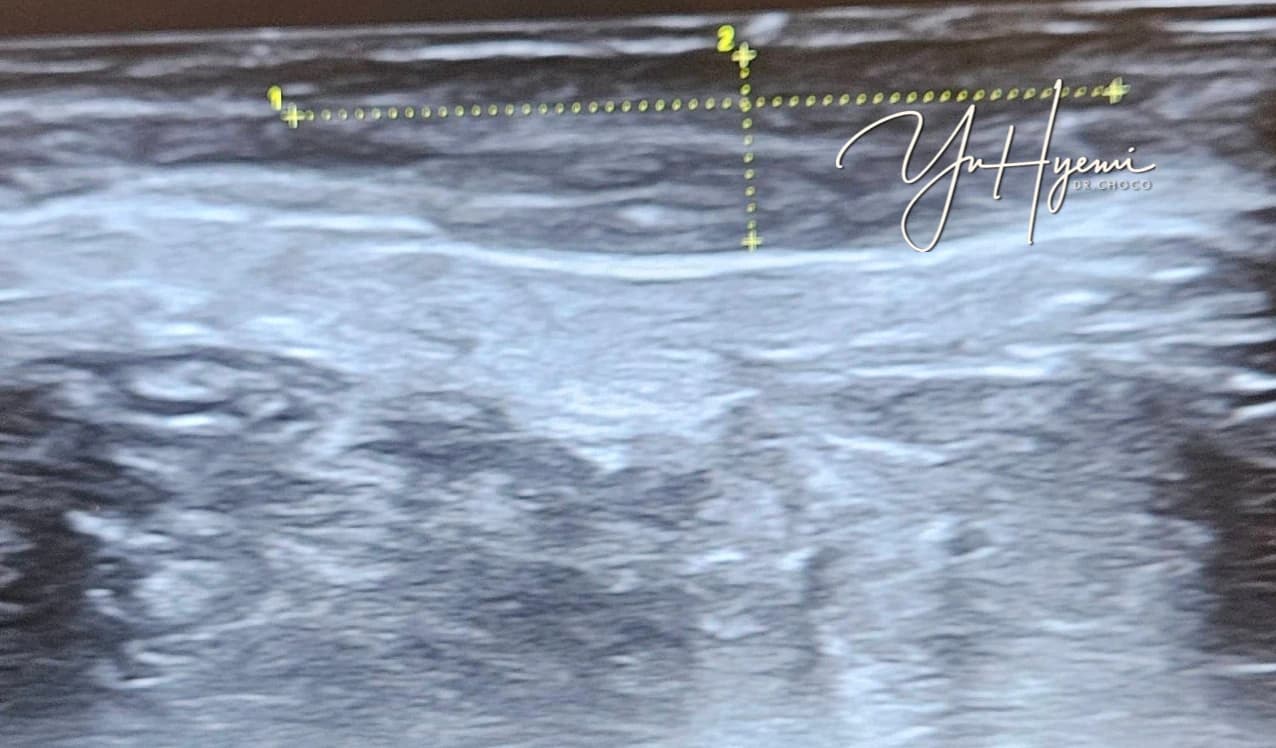
What the source ultrasound showed
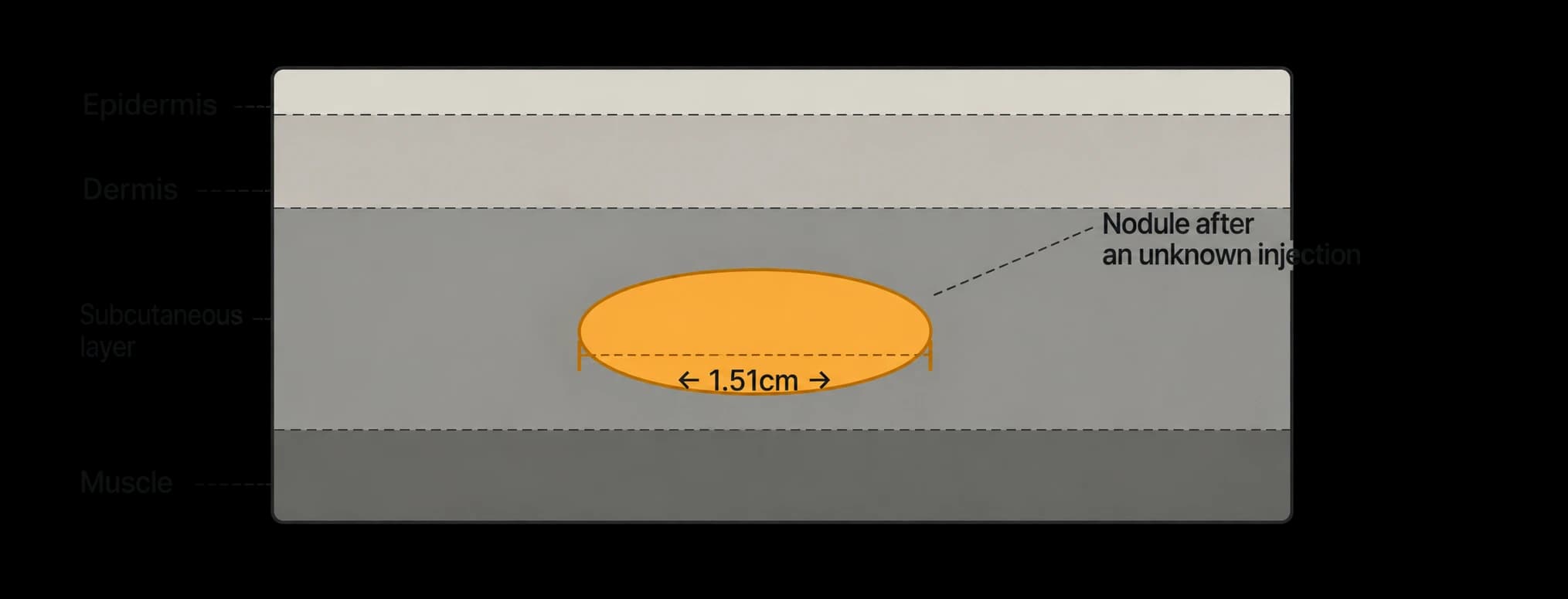
Source caption: a superficial bright lesion measured approximately 1.51cm by 0.54cm. Brightness and dimensions do not identify the injected substance or establish that every similar lesion will persist for years.
Why knowing the material matters
The source cites Modarressi et al., 2020, describing 26 patients with post-filler nodules.
The source quotes 65.4% as having an unknown material before histopathological analysis. This is a finding in selected cases, not all filler patients.
Material identification can change treatment choices. Hyaluronidase acts on HA, while inflammatory reactions to other materials require different assessment. With unknown products, history, examination, imaging, response and sometimes tissue sampling may all be relevant.
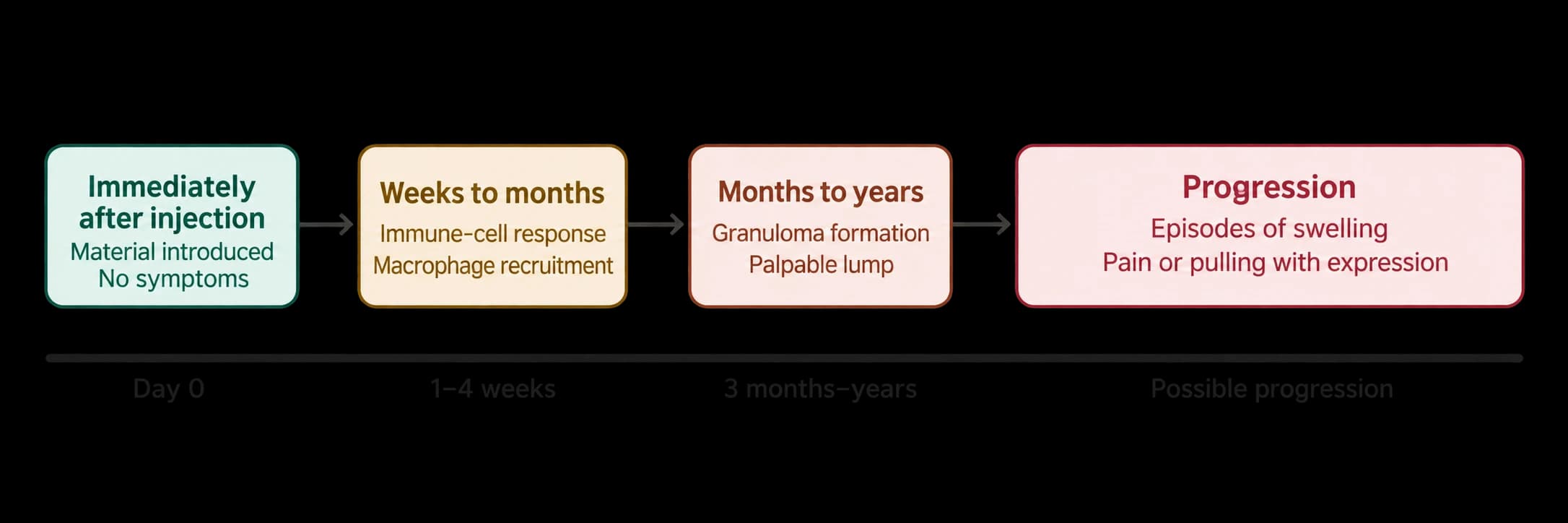
Delayed reactions can occur
A lack of an immediate reaction does not exclude later problems. The source cites a 2017 Korean radiology paper with 39 patients, discussing delayed foreign-body reactions.
The source describes granulomas arising months to years later and several proposed mechanisms, including immune responses and contamination. These mechanisms are not all established in an individual lesion.
A new lump long after treatment needs assessment, including a careful record of earlier products and sites.
Limits of complete removal
A 2015 review by Lee and Kim discusses removal of foreign-body granulomas.
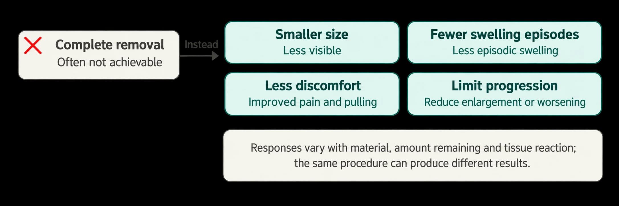
The source notes that poorly defined borders can make removal incomplete and that surgery may leave scarring or deformity.
This possibility does not mean that every nodule is permanent or cannot improve.
Treatment goals
Reducing pain, recurrent swelling and interference with expression can be meaningful even when complete removal is not possible. The source compares this with improving function after an injury rather than expecting a perfect return to the original state.
What ultrasound can and cannot do
History and examination remain important. Ultrasound can help assess deposits and surrounding structures but cannot always identify the substance or confirm or exclude infection.
The source cites Mlosek et al., Journal of Ultrasonography, 2021, on distinguishing granulomas and filler deposits using high-frequency ultrasound.
A deposit, an inflammatory granuloma and infection are different problems. Imaging supports evaluation rather than replacing the clinical diagnosis.
Options reported in the literature
The source cites Cerón Bohórquez and Desyatnikova’s 2024 PCL granuloma case.
The source emphasizes that delayed reactions can have multiple contributing factors.
Reported approaches include hyaluronidase for HA, anti-inflammatory medication for selected granulomas, infection assessment and treatment, and surgery when appropriate. Unknown substances may require staged assessment or biopsy. These are clinician-led options, not a self-injection protocol.
Assessment may be appropriate for recurrent swelling, pain or pulling with expression, increasing size, a new delayed lump or tenderness.
This article introduces literature and does not replace individual care.
References cited by the source
Modarressi A, Nizet C, Lombardi T. Granulomas and nongranulomatous nodules after filler injection. JPRAS. 2020. https://pubmed.ncbi.nlm.nih.gov/32928687/
Kim JH, et al. Ultrasonographic Features of Complications Associated with Injectable Fillers in the Cervicofacial Region. Journal of the Korean Society of Radiology. 2017;77(4). https://jksronline.org/DOIx.php?id=10.3348/jksr.2017.77.4.222
Lee JM, Kim YJ. Foreign body granulomas after the use of dermal fillers. Archives of Plastic Surgery. 2015;42:232–239. https://pubmed.ncbi.nlm.nih.gov/25798398/
Cerón Bohórquez JM, Desyatnikova S. Ultrasound-guided Treatment of Polycaprolactone Granuloma. PRS Global Open. 2024;12:e5610. https://pmc.ncbi.nlm.nih.gov/articles/PMC10860983/
Source: Original post on Dr. Hyemi Yu’s Naver blog (Korean)
The original publication date and the website revision date are shown separately.