
JOURNAL
Persistent under-eye nodules after dissolving injections: ultrasound assessment
An individual under-eye nodule case and the interpretation of ultrasound, Doppler and cited literature.
These are ultrasound and Doppler observations in an individual case. Brightness or flow signals alone cannot establish material, fibrosis, causation or ESWT efficacy. Laterality, case-image consent and the applicability of cited studies require review.
“I have had dissolving injections several times elsewhere, but I can still feel the lump.” This is a concern some patients bring to consultation.
A history of under-eye filler, PN, Sculptra or cheekbone surgery
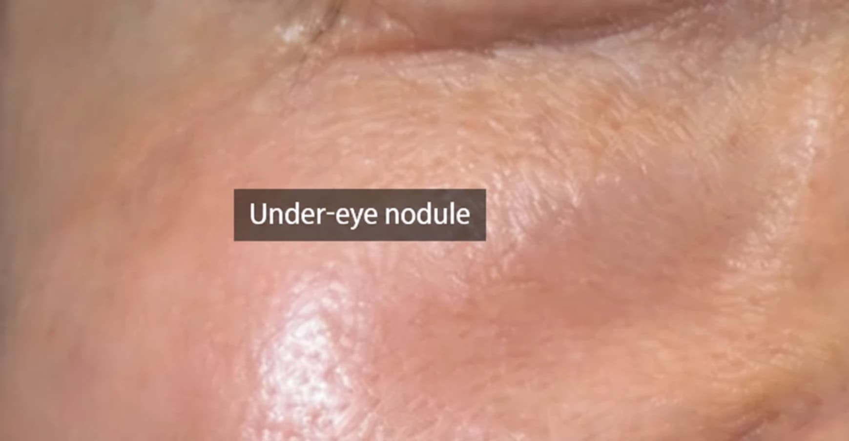
A patient may describe repeated hyaluronidase injections while under-eye lumps persist and the face feels heavier or less expressive. The source discusses the possibility of different materials and tissue reactions being present together, rather than assume everything is dissolvable HA.
Under-eye nodules discussed after Sculptra or Juvelook
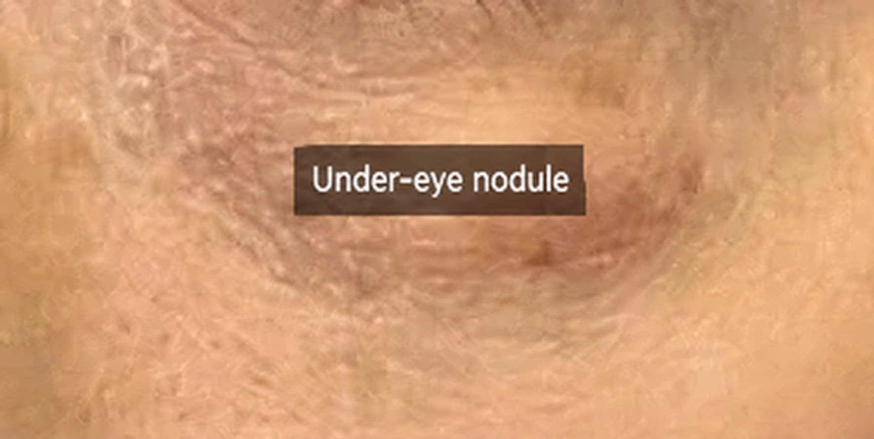
The case history described repeated Sculptra (PLLA) around the cheek, Rejuran Eye (PN) under the eyes, other filler procedures and cheekbone surgery approximately 15 years earlier. The aim had been to address under-eye hollowness.
The author describes a later appearance with volume but heaviness and reduced expression, sometimes called “puffy.” Previous dissolving procedures had not resolved the concern. Under-eye findings were prioritized. The case narrative’s laterality requires review against the original records.
Looking beneath the surface
Palpation and ultrasound provide different information. The source compares listening through a wall with having a view of what lies behind it.
Source image 1: before treatment, left under-eye (Lt lid)
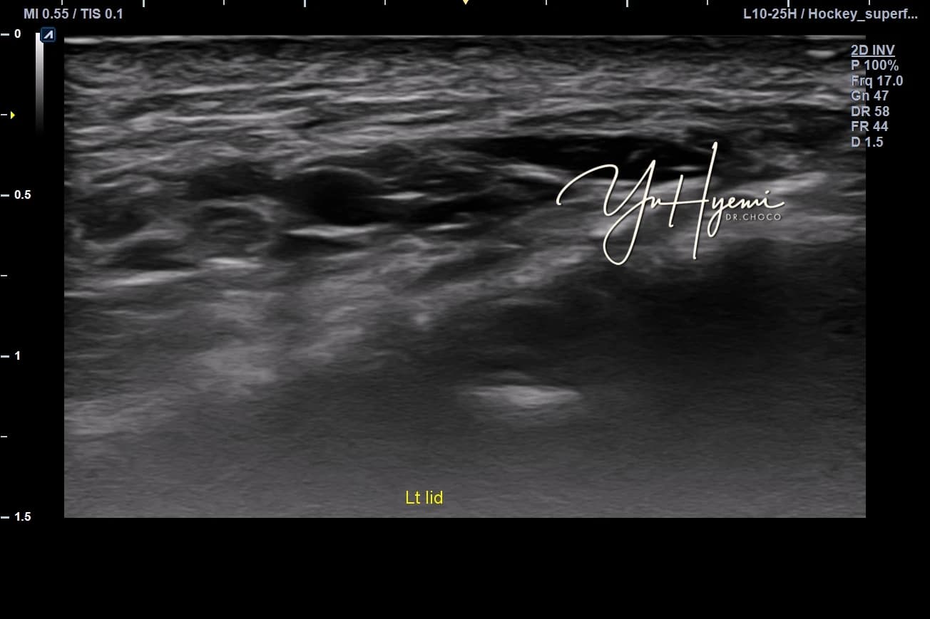
The author describes multiple irregular, poorly defined hypoechoic findings in subcutaneous tissue, intermingled with surrounding structures. The source considers prior biostimulators or mixed materials as possible explanations.
An ultrasound appearance alone cannot identify the injected material or prove fibrosis. The original post’s explanation for why hyaluronidase did not resolve the finding is a clinical interpretation requiring review.
Hyaluronidase is used for HA-related filler issues; it is not a universal dissolver of fibrotic tissue or PLLA. The source refers to a JMIR Dermatology review.
Kroumpouzos G et al. JMIR Dermatol. 2024;7:e50403.
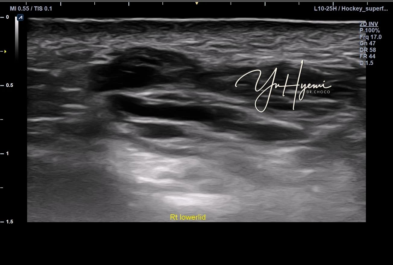
Source image 4: before treatment, right lower eyelid
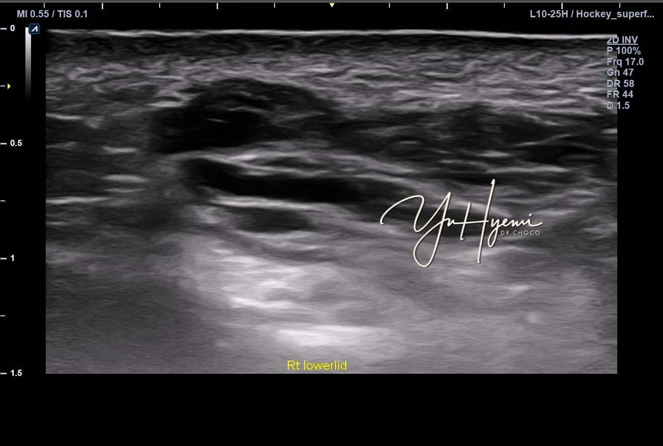
The source describes multiple small, hypoechoic, honeycomb-like findings on the right as well. It reports marked protrusion on that side and prioritization for treatment. Left/right descriptions should be verified against image labels.
Doppler observations around ESWT
The source discusses blood-flow assessment in fibrotic tissue and proposes a relationship between circulation and recovery. Its general statement that fibrosis means blood flow is blocked should not be treated as a diagnostic rule.
Source image 2: Doppler before ESWT, as labeled in the original case
The author describes limited color-flow signals around the finding, with small blue and red areas. A further image follows treatment.
Source image 3: Doppler after ESWT (Post ESWT rt tt)
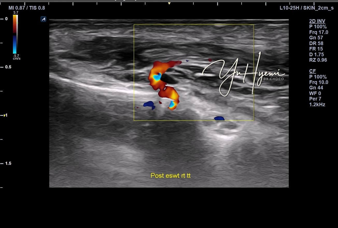
On re-examination after extracorporeal shock-wave therapy, the author describes smaller-appearing areas and more orange/red signals around the finding. This is an individual imaging observation, not proof that a material was fragmented or that circulation was restored by treatment. Settings and assessment conditions also require review.
The source refers to literature examining growth factors, blood/lymphatic responses, fibrosis and ECM remodeling after ESWT. Applicability depends on the study context.
It cites a report discussing deep dermal/subdermal fibrosis and nodules after PLLA, with ultrasound observations before and after low-energy ESWT.
Ko J, Cho SB. Skin Res Technol. 2024;30:e70082.
The source also discusses molecular findings involving TGF-β1, α-SMA, collagen I and fibronectin. These experimental findings do not by themselves establish an individual treatment effect.
The author uses this as an analogy for slowing fibrotic activity.
Reference discussed: PMC5796073, concerning fibrosis-related molecules in hypertrophic-scar fibroblasts.
Questions this case may raise
The author addresses people concerned about increasing heaviness after repeated filler or biostimulator procedures, persistent lumps after dissolving treatment, or concerns that have only been assessed by touch.
Complications need not be a source of shame. Identifying what may be present and where it lies can inform assessment, but this case does not establish one treatment for all such concerns.
This article shares literature and clinical experience without guaranteeing a procedure’s effects. Individual suitability requires specialist assessment.
References cited in the source
- Kroumpouzos G, Treacy P. Hyaluronidase for Dermal Filler Complications. JMIR Dermatol. 2024;7:e50403. PubMed.
- Ko J, Cho SB. Clinical Efficacy and Safety of Low-Energy ESWT for Various Conditions of Deep Dermal and Subdermal Fibrosis. Skin Res Technol. 2024;30:e70082. PMC11452247.
- ESWT and fibrosis-related molecules in hypertrophic-scar fibroblasts. PMC5796073.
Study populations, indications, exact descriptions and causal interpretations require medical review.
References linked by the source
Source: Original post on Dr. Hyemi Yu’s Naver blog (Korean)
The original publication date and the website revision date are shown separately.