
JOURNAL
Recurrent PDLLA nodules and treatment limits
Recurrent PDLLA nodules and treatment limits. Source observations, research context and limitations.
This article discusses the source author’s earlier literature, cases and experiences. Research populations and limitations matter; individual diagnosis and treatment require clinical assessment.
A clinical account of recurrent PDLLA nodules
This article records an individual clinical course and the author’s perspective. Publication of the original blog does not establish approval of all medical claims or of this republication.
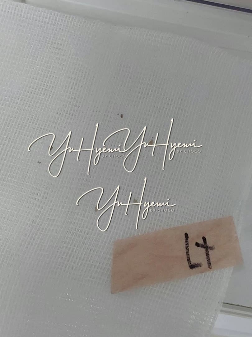
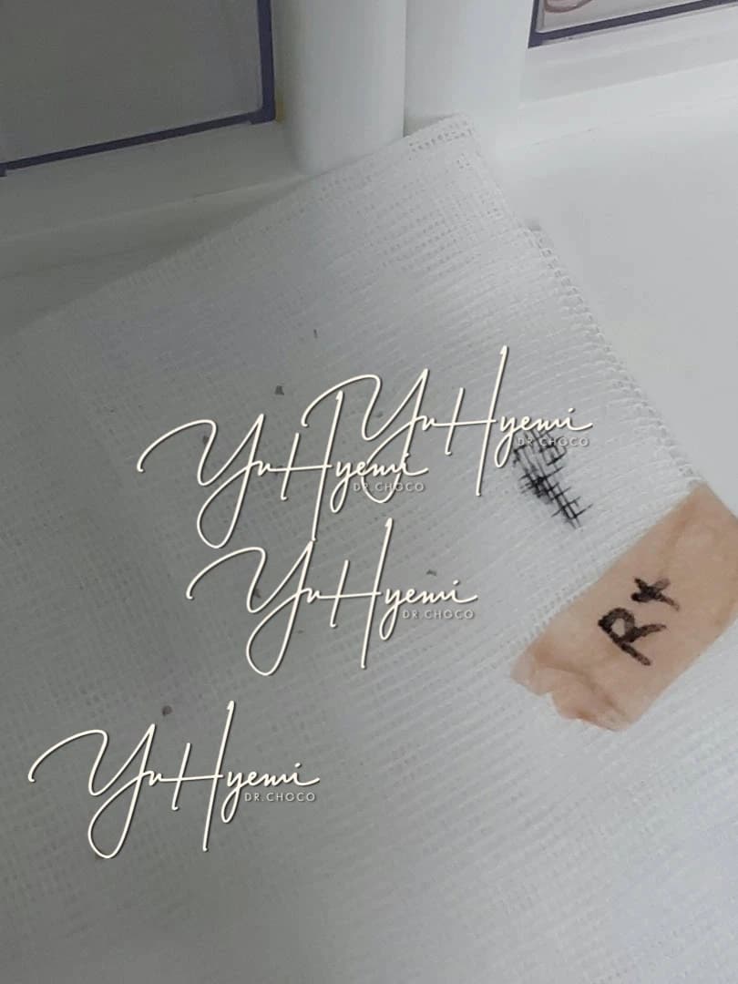
The source describes more than 20 nodules along the neck after treatment elsewhere. Medication injections, an RF needle and laser-based approaches were reported to reduce their size, but residual lesions remained visible in thin skin and caused distress. The author discusses recurrence after apparent improvement rather than a uniform pattern for all PDLLA nodules.
The author considered the lesion and its surrounding tissue, including ultrasound findings, circulation and fibrosis. This individualized approach is a clinical perspective rather than proof of a particular functional-medicine protocol.
Recurrence after apparent reduction
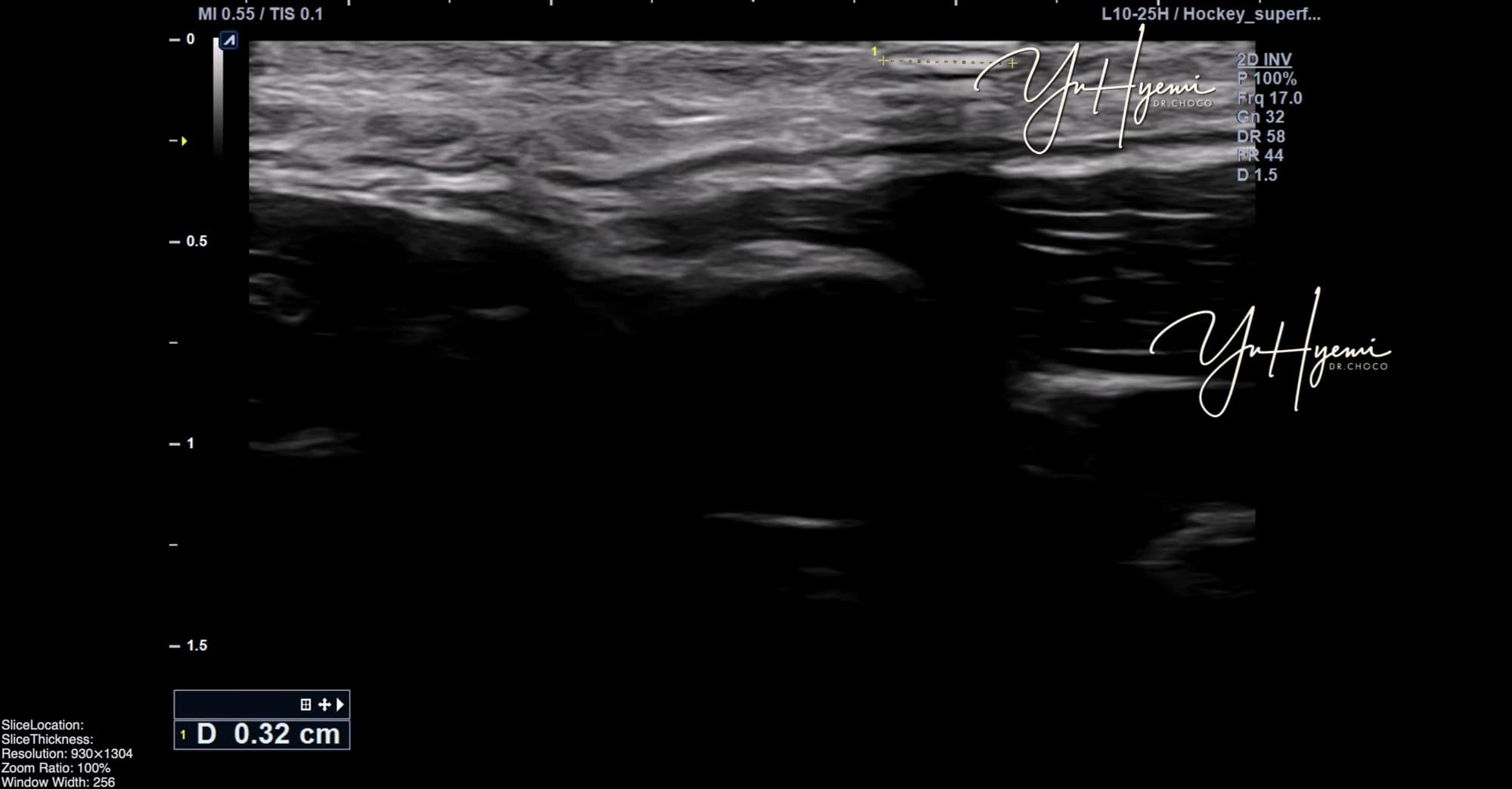
The source reports one measurement changing from 0.32cm to 0.08cm before renewed enlargement. PDLLA-related collagen responses were proposed as one explanation. Thin skin may make small lesions noticeable, but ultrasound brightness does not by itself prove tissue stiffness, composition or the cause of recurrence.
Source caption: a hyperechoic lesion measured at 0.32cm.
Source caption: a smaller residual lesion after several injections.
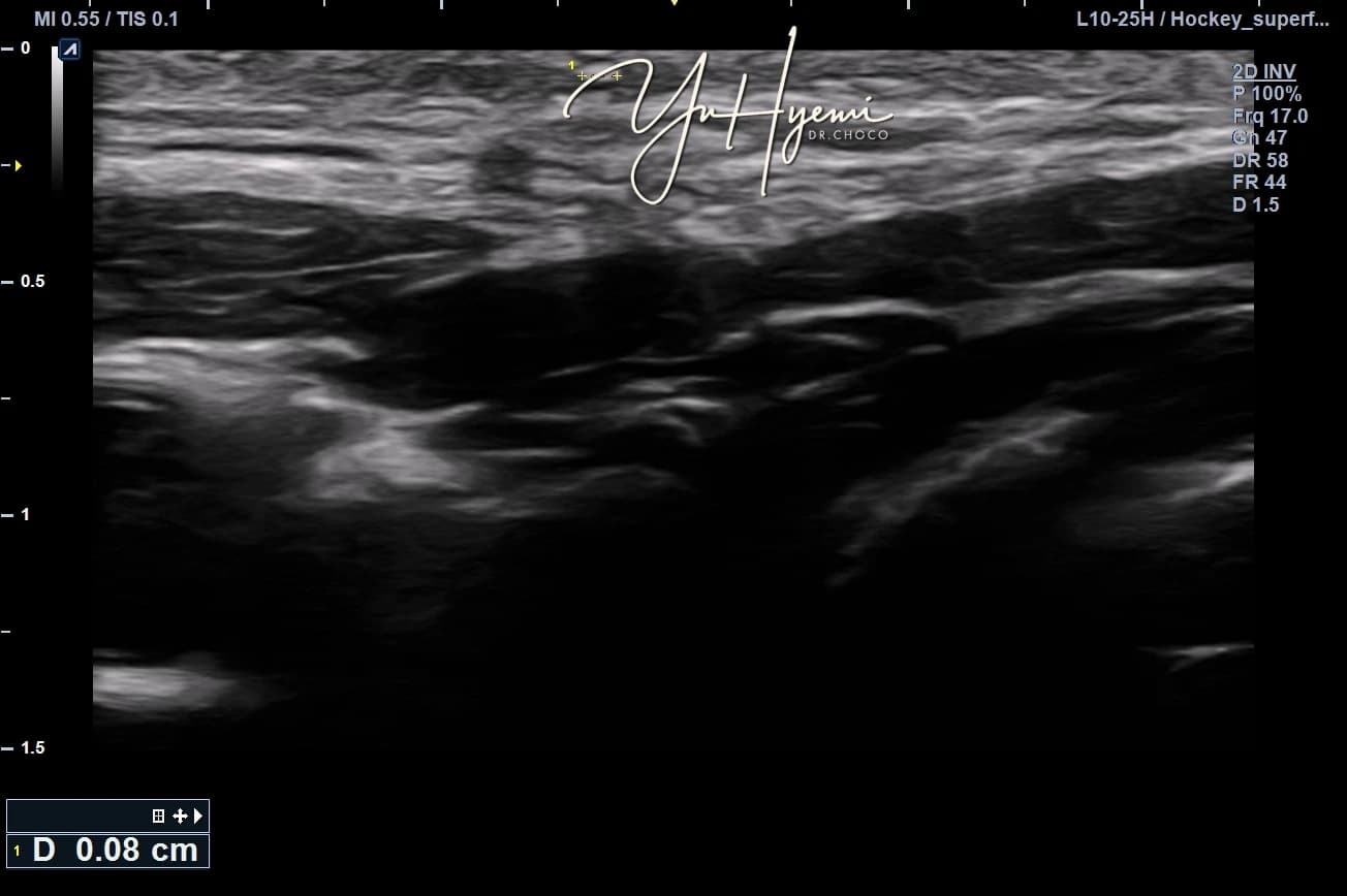
The author describes reduced measurements and a less distinct boundary, while a small lesion remained visible. Recurrence prompted reassessment. An ultrasound impression cannot independently demonstrate restored elasticity or prove that residual PDLLA was producing new collagen.
Reassessing diagnosis and options
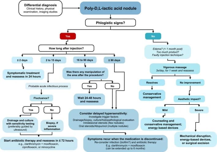
The source records that some persistent lesions were removed by the clinician after earlier injection and energy-based treatments. Detailed needle curettage, cutting and suturing instructions were omitted from this patient version. Removal may involve scarring, infection, injury and incomplete clearance; diagnosis and possible histopathology require assessment. Claims that heat or massage easily reshapes PDLLA are not treated as a clinical standard.
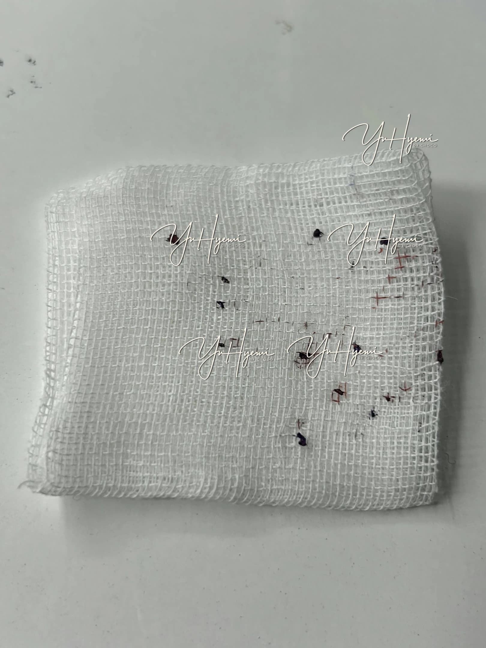
The author reports satisfaction in an individual outcome and a staged wound-care course, but this is not a recommendation for every nodule to be removed. The source then discusses keratinocytes, dehydration, TGF-β and IL-6 as possible contributors to fibrosis. These mechanisms arise from different settings and do not prove that dry skin caused this lesion or that a hydration-focused treatment prevents recurrence.
Treatment choices also depend on the patient’s circumstances, recovery time and tolerance of risk. The author’s emphasis is to understand the person and tissue rather than simply chase every palpable lump. Neither surgery nor conservative treatment has a guaranteed course or outcome in this account.
Source: Original post on Dr. Hyemi Yu’s Naver blog (Korean)
The original publication date and the website revision date are shown separately.