
JOURNAL
Ultrasound and lessons from a third anatomy workshop
Reflections on a third ultrasound anatomy workshop and earlier research activity.
This records a workshop and earlier publications. Publication/citation details, roles and image rights require verification; ultrasound does not remove every procedural risk.
After filler treatment, someone may notice asymmetry, a firm lump or swelling lasting longer than expected and wonder whether reassurance alone is enough. Such concerns deserve assessment rather than an assumption from the passage of time.
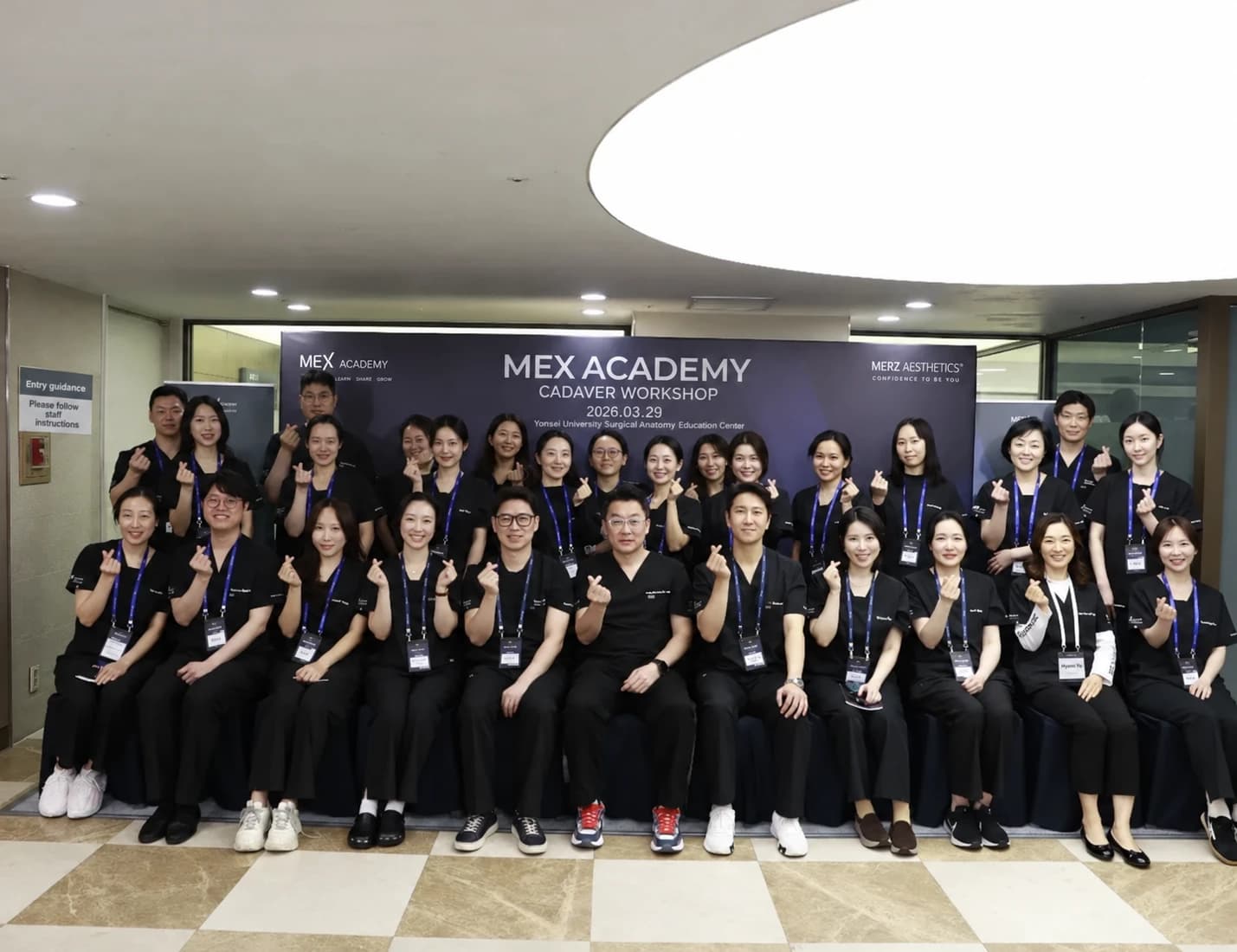
I recently completed my third ultrasound-guided anatomy workshop. It was a MEX Academy cadaver workshop associated with Merz, whose portfolio includes Radiesse, Ultherapy and Xeomin. This time I participated as faculty for the first time.
During my first anatomy workshop the previous year, I could see structures but struggled to connect them with textbook knowledge. Repeated workshops and clinical ultrasound experience have helped me understand needle position, layers and vessels more spatially.
It is rather like the difference between memorizing a map and walking a route many times. This is my account of learning, not proof that imaging removes all procedural risk.
Professor Hee-Jin Kim provided ultrasound-guidance teaching during approximately nine hours of anatomical dissection.
The source recalls a message that patients increasingly seek minimally invasive procedures, making careful ultrasound interpretation important. The exact quoted wording requires confirmation.
With open surgery, tissue can be viewed directly; with a needle-based procedure, other sources of anatomical information are needed. The original uses a dark-room analogy to emphasize this challenge.
It is the author’s perspective on the value of ultrasound, not evidence that every procedure performed without it is unsafe or that imaging guarantees safety.
The source describes research and publications involving ultrasound-guided assessment and treatment. It states that two papers published in the preceding year concerned ultrasound-guided hydrodissection and nerve-related treatment. Exact publication details and the scope of the research require verification.
The original post refers to a journal impact factor of 3.3. Journal metrics do not establish the effectiveness of an individual treatment.
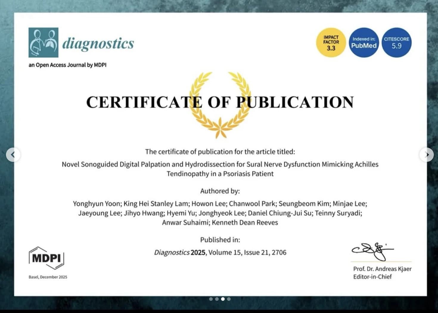
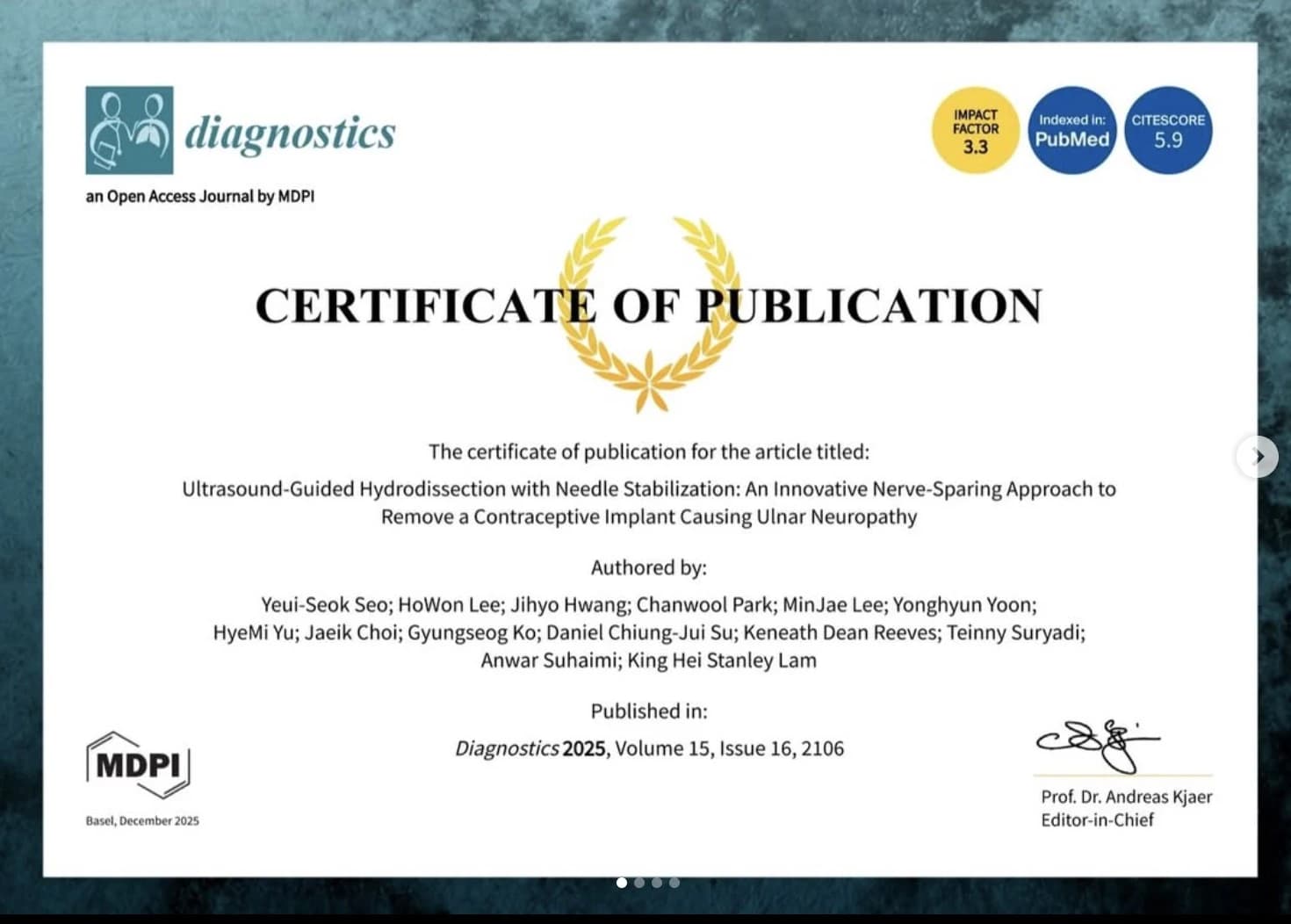
Writing and publishing can provide a structured way to report and scrutinize observations. Publication alone, however, does not prove a treatment works for every indication.
The source describes concerns in people previously treated elsewhere, including uncertainty about material location. Ultrasound may add information about tissue, surrounding vessels or an area requiring further assessment. These descriptions do not establish a universal diagnosis, causation or superiority over other providers.
Workshop participation as faculty.
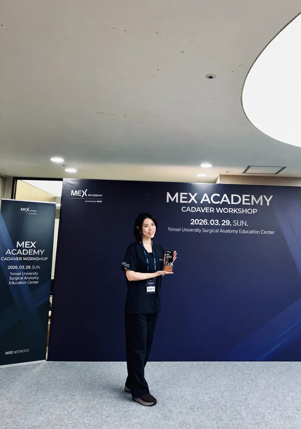
With a colleague identified in the original as Dr. Jimin Lee of Director Dermatology; official English affiliation and name require confirmation.
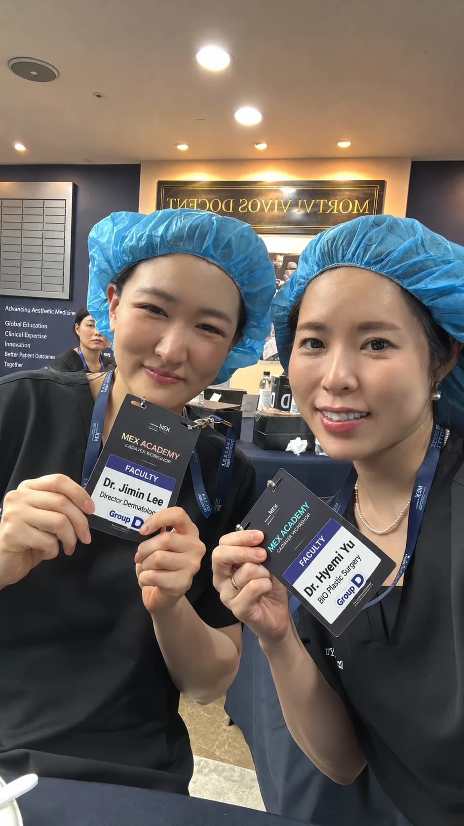
The workshop deepened my anatomical understanding and reminded me why I keep studying: to give more considered answers to people concerned about complications.
If something seems unusual after a procedure, assessment can help establish what is present and where. Whether ultrasound or another investigation is appropriate depends on the clinical situation.
This article records participation in MEX Academy/IARM cadaver workshops and research activity related to ultrasound-guided nerve treatment. Historical roles, affiliations and publication details require clinic review.
Source: Original post on Dr. Hyemi Yu’s Naver blog (Korean)
The original publication date and the website revision date are shown separately.